
Melbourne — Orthopaedics
Cartilage Damage in the Hip:
Causes, Symptoms & Treatment
We explain how hip cartilage damage occurs, the symptoms to watch for, how it is diagnosed, and the most effective non-operative and surgical treatments—so you can return to work, sport, and daily life with confidence.

What Is Hip Cartilage Damage?
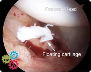
The hip’s ball (femoral head) and socket (acetabulum) are covered by articular cartilage, a smooth, low-friction layer that cushions and protects the joint. Cartilage damage (also called chondral injury or chondral defect) occurs when this lining softens, frays, or wears through. Because cartilage has limited blood supply, it heals poorly without targeted treatment.
Common Causes of Hip Cartilage Damage
- Femoroacetabular Impingement (FAI): extra bone on the ball or socket causes abnormal contact during movement, leading to progressive cartilage wear. See FAI overview.
- Labral tears: a torn labrum destabilises the socket seal, increasing shear forces on cartilage. See Labral tear of the hip.
- Trauma or sports injury: falls, twists, or dislocation can cause focal chondral defects.
- Early osteoarthritis: cumulative wear results in thinning and fissuring of cartilage.
- Inflammatory arthritis: immune-mediated inflammation can erode cartilage.
- Ligamentum teres pathology: instability or tears may overload cartilage; see Ligamentum teres tears.
Symptoms
- Deep groin pain (sometimes buttock or lateral hip pain)
- Stiffness and reduced range of motion—especially with twisting or pivoting
- Clicking, catching, or locking within the joint
- Pain after sitting, driving, or sport; relief with rest
- Occasional referral to the thigh or knee
How We Diagnose Hip Cartilage Damage
Diagnosis combines clinical assessment with targeted imaging:
- Specialist examination: movement tests (e.g., FADIR) identify impingement and instability patterns.
- Weight-bearing X-rays: assess bony morphology and signs of arthritis.
- MRI : visualises cartilage, labrum, bone marrow oedema, and associated pathology.
- Hip arthroscopy: minimally invasive procedure that directly visualises cartilage and treats confirmed lesions. See Hip arthroscopy (keyhole surgery).

Treatment Options
Non-operative Care
- Activity modification: reduce deep flexion and pivoting while symptoms settle.
- Physiotherapy: targeted strengthening (gluteal/core), mobility, and neuromuscular control to offload the joint. See our Physiotherapy service.
- Medications: short courses of anti-inflammatories as appropriate.
- Image-guided injections: corticosteroid for short-term inflammation control; biologic options (e.g., PRP) may be considered on a case-by-case basis.
Surgical Care (Hip Arthroscopy)
When symptoms persist or imaging confirms significant defects, we may recommend hip arthroscopy to treat the root cause and address the cartilage directly.
- FAI correction (osteoplasty): reshaping the ball and/or socket to remove impingement.
- Labral repair: restores the suction seal and stability.
- Cartilage-focused techniques:
- Chondroplasty (debridement): smooths unstable flaps to reduce catching.
- Microfracture: stimulates fibrocartilage fill for small, contained defects.
- Hip replacement: in advanced, diffuse loss, hip replacement may be the most predictable option for pain relief and function.
| Treatment | Best for | Aim | Recovery guide |
|---|---|---|---|
| Physiotherapy | Mild symptoms, early changes | Reduce pain, improve stability | 6–12 weeks structured program |
| Injection therapy | Inflammation flares, diagnostic clarification | Short-term symptom control | Days to 2–6 weeks effect (varies) |
| Arthroscopy + chondroplasty | Unstable flaps/catching | Smooth surfaces, reduce mechanical symptoms | Return to office work ~1–2 weeks; sport later |
| Microfracture | Small, contained defects | Stimulate defect fill | Protected loading 4–6 weeks; rehab 3–6 months |
| Hip replacement | Diffuse, advanced loss | Reliable pain relief & function | Hospital stay protocols vary; rehab 6–12 weeks+ |
Recovery & Prognosis
Outcomes depend on defect size, location, and whether underlying causes (such as FAI or labral tears) are corrected. Early, targeted treatment can reduce pain, restore function, and slow progression to osteoarthritis. Larger, diffuse lesions and long-standing symptoms carry a higher risk of requiring joint replacement later.
We provide a staged rehabilitation plan with objective milestones, clear activity progressions, and communication with your physiotherapist to keep recovery on track.
Appointments & Second Opinions
If you have persistent hip pain, stiffness, or clicking that hasn’t improved after six weeks, we can help with accurate diagnosis and a tailored plan.
Frequently Asked Questions
Is cartilage damage the same as arthritis?
Cartilage damage refers to a focal defect or early wear. Osteoarthritis implies more widespread, progressive loss of cartilage with joint changes such as bone spurs and narrowing on X-ray.
Can hip cartilage damage heal on its own?
Cartilage has a very limited capacity to heal spontaneously. Symptoms may settle with rest and physiotherapy, but persistent mechanical symptoms or instability often need targeted treatment.
When is hip arthroscopy recommended?
When non-operative care fails and imaging or examination confirms a treatable cause (e.g., FAI, labral tear, focal chondral defect) that aligns with your symptoms and goals.
How long is recovery after microfracture?
Typically protected weight-bearing for 4–6 weeks, with progressive strengthening thereafter. Return to running and pivoting sports is later and personalised.
Will I eventually need a hip replacement?
Many people improve with joint-preserving strategies. If cartilage loss becomes diffuse and advanced, hip replacement may provide the most predictable pain relief and function.
